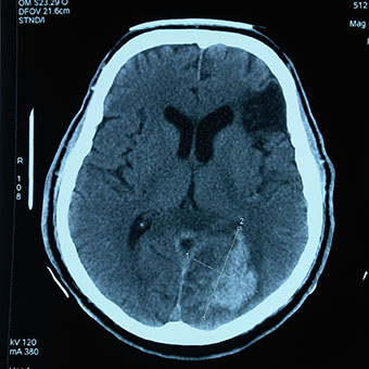
Computerised tomography
CT stands for computerised tomography. A CT scanner uses x-rays and a computer to create detailed images of the inside of the body. It's good at visualising the areas which do not show so well on ordinary x-ray images. CT scan images are used to detect and diagnose medical conditions and injuries in different parts of the body, including the head and abdomen. Therapeutic radiographers (also known as radiotherapists) use CT to plan and verify patients' treatment.
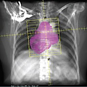
Radiotherapy treatment
Therapeutic radiographers use hi-tech machines to treat people who have cancer. Called a linear accelerator, or linac for short, the machine generates x-rays for radiation therapy (radiotherapy). The image below (courtesy of The London Clinic) shows an image which is used for planning radiation treatment.
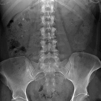
X-ray
X-rays are a type of radiation that can be used to look into your body without surgery. The x-rays are photons of energy which are absorbed at different rates by the body and it's this pattern that creates the x-ray image. X-rays were discovered by Wilhelm Conrad Röntgen in 1895 and the first x-ray taken was of his wife's hand, which clearly showed her bones and ring. X-rays are also used to treat cancer. This is known as radiation therapy or radiotherapy.
Patient care
Caring for patients is at the heart of the radiographer’s role. Good communication skills and a compassionate approach are essential qualities for every radiographer.
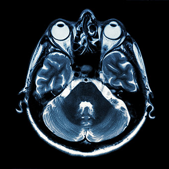
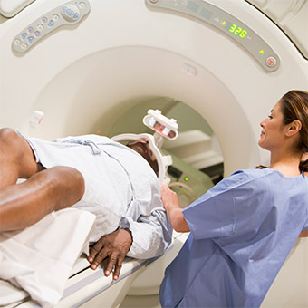
MRI
MRI is short for Magnetic Resonance Imaging. This machine uses strong magnetic fields and radiowaves to form images of the body. An MRI machine does not use x-rays to produce an image and it can examine almost any part of the body. However, because of its strong magnet, not all patients can undergo an MRI scan – people with pacemakers, for example.
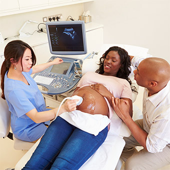
Ultrasonography
Soundwaves can be used to visualise body structures such as tendons, muscles, joints, vessels and internal organs. It’s most often used to examine unborn babies. The person who does this is also known as a sonographer or ultrasonographer.
Radionuclide imaging
Nuclear medicine uses radioactive isotopes to diagnose and treat patients. It records the radiation emitted from within the body to show how the body is functioning. Nuclear medicine is a term that encompasses several different imaging techniques. These include positron emission tomography (PET) which is used with CT (PET-CT) and single photon emission computed tomography (SPECT).
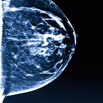
Mammography
Low energy x-rays are used to take an image of a patient’s breasts. Mammography is used to screen for, and diagnose, breast cancer. The person who does this is also called a mammographer.